应用离子对HPLC?ELSD法测定磷霉素钠中磷霉素及有关物质的含量
【摘要】 建立了HPLC?ELSD法测定磷霉素钠中磷霉素及有关物质的含量。采用ODS柱,柱温35℃,以0.015mol/L正辛胺溶液(用冰乙酸调节pH至5.2)∶乙腈(90∶10)为流动相,蒸发光散射检测器温度为45℃,雾化气体压力为0.35MPa。磷霉素及其二醇物分别在26.0~207.7和7.6~60.6μg/ml范围内呈良好的线性关系,两者的定量限分别为8.7和7.7μg/ml。
【关键词】 离子对HPLC?ELSD 含量测定 磷霉素钠 有关物质
Fosfomycin is a synthetic, broad?spectrum bactericidal antibiotic for oral administration. It has in vitro activity against a wide range of Gram?positive and Gram?negative aerobic microorganisms, which are associated with uncomplicated urinary tract infection[1]. The fact that fosfomycin has no UV?Vis absorption has led to the development of few analytical methods for its determination. As far as we know, only the HPLC with refractive index (RI) detection method, i.e. the British Pharmacopoeia (BP) method used for analysis of fosfomycin trometamol[2a] can provide separation for fosfomycin and related substances. Due to the poor sensitivity of RI detector, very large amount of sample is injected which causes the fosfomycin peak to be broadened, and the resolution between fosfomycin and related substances might be reduced. Furthermore, due to the poor buffer capacity of the mobile phase and least stability of the NH2 column, the BP method is not robust enough. According to BP[2b], fosfomycin sodium and disodium 1,2?(dihydroxypropyl) phosphonate (diol substance) are determined by titrimetric method which lacks of accuracy. Utilizing the Chinese Pharmacopoeia (ChP) method[3a], fosfomycin in fosfomycin sodium is determined by microbiological analysis, while fosfomycin sodium diol substances are determined by titrimetric method. This article provided an ion?pair HPLC?ELSD method for the determination of fosfomycin and related substances in fosfomycin sodium.
1 Experiment
1.1 Materials
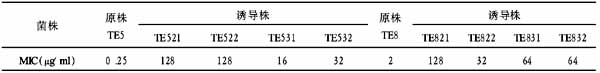
Fosfomycin sodium and fosfomycin calcium were provided by Shanghai Asia Pioneer Pharmaceuticals Co., Ltd. Fosfomycin?Chinese National Chemical Reference Standard (CRS) of activity 651μg/mg, fosfomycin trometamol?European Pharmacopoeia CRS of activity 53.1%, and fosfomycin trometamol impurity A (diol substance)?European Pharmacopoeia CRS were used. Octylamine was purchased from Aldrich. Acetontrile was HPLC grade and obtained from E. Merck. All other chemicals were analytical grade reagents. All solutions were prepared with distilled water for HPLC.
1.2 Instrumentation
The elution was performed with an Agilent 1100 HPLC system. A Sedex 75 evaporative light?scattering detector was used for detection.
1.3 Procedures
The mobile phase contained 0.015mol/L octylamine solution (adjusted to pH5.2 with glacial acetic acid)∶acetonitrile (90∶10) and the flow rate was maintained at 1.5ml/min. Chromatographic separations were performed at 35℃ on a 250mm×4.6mm i.d. column packed with 5μm Luna 5μ C18 (2) and the injection volume was 50μl. The detector was set at temperature 45℃, the gain 8 and the pressure of air 0.35MPa.
1.4 Solution preparation
A 0.97 mg/ml fosfomycin trometamol CRS stock solution was prepared in mobile phase and stored with protection from light in the refrigerator. Working standard solutions in the range 26.0~207.7μg/ml of fosfomycin were freshly prepared in mobile phase. Sample solution used for assay was 0.1mg/ml in mobile phase. For fosfomycin sodium diol substances calibration, a 0.42mg/ml fosfomycin trometamol impurity A CRS stock solution was prepared in mobile phase and stored with protection from light in the refrigerator. Working standard solutions in the range equivalent to 7.6~60.6μg/ml of disodium 1,2?(dihydroxypropyl) phosphonate were freshly prepared in mobile phase. Sample solution used for impurity detection was 5.5mg/ml in mobile phase.
2 Results and discussion
2.1 Method development and optimization
Fosfomycin and its related substances all possess negative charge in aqueous solution. Due to their relati?vely hydrophilic properties, they are eluted near the dead volume in a typical reversed phase HPLC system. For this reason, an ion?pair reagent might be added to the mobile phase to form, with the ionic analytes, ion?pairs, which according to their hydrophobicity, will be retained on the reversed stationary phase.
ELSD is described as a quasi?universal detection mode suitable for non?absorbing analytes. The response does not depend on the solute optical properties, any compound less volatile than the mobile phase could be detected. The detector response shows a double logarithmic relationship between this signal and the analyte concentration. Its ability to perform quantitation of substances with lack of standard material, as it shows nearly equal response factors for molecules with about equal molecular mass and similar structure, is well?established.
Since ELSD demands the evaporation of the mobile phase prior to light?scattering step, mobile phase of high volatility are required. Octylamine and acetic acid, which is used to adjust the pH of mobile phase and to provide buffer capacity for the mobile phase, are applicable.
The experimental results indicated that at pH (5.2±0.2) and 6%~10% acetonitrile content in the mobile phase, fosfomycin and its related substances could be well separated from each other. Addition of acetonitrile to the mobile phase resulted in decreased retention of all analytes. Maintaining the pH of buffer at 5.2, as the concentration of octylamnine was increased from 0.005mol/L to 0.020mol/L, the retention time of all analytes increased. Further increasing the concentration of octylamnine increased the baseline noise significantly.
The optimized mobile phase contained 0.015mol/L octylamine solution (adjusted to pH5.2 with glacial acetic acid)∶acetonitrile (90∶10). A particle of CH3(CH2)7NH3+?fosfomycin will be formed during desolvation and subsequently detected by light scattering. In the case of sodium, Na+?CH3COO- is formed under these conditions and subsequently detected.
The separation of fosfomycin and related substances was illustrated in Fig.1, which shows baseline separation between fosfomycin and related substances. Sodium was eluted near the dead volume due to its possessing positive charge. Fig.2 shows chromatogram for a sample solution used in assay. The theoretical plate number and the tailing factor of the fosfomycin peak were around 18,000 and 1.08 respectively (Fig.2).
The chromatogram in Fig.3 indicates that this method can probably be applied to determination of fosfomycin and related substances in fosfomycin calcium. According to BP[2c], fosfomycin calcium and calcium 1,2?(dihydroxypropyl) phosphonate (diol substance) are determined by titrimetric method, while using the ChP method[3b], fosfomycin in fosfomycin calcium is determined by microbiological analysis, and fosfomycin calcium diol substances are determined by titrimetric method. Due to its relatively lower solubility in the mobile phase, the concentration of sample was decreased, and 100μl was used as compromising injection volume.
2.2 Method validation
A series of working standard solutions (26.0~207.7μg/ml of fosfomycin) were analyzed in duplicate and the peak areas (A) were used to construct the logarithmic calibration curve. The regression equation and the correlation coefficient (r) were logA=1.5898logC+1.9142, and 0.9991 (n=7) respectively. For fosfomycin sodium diol substances, a series of working standard solutions (equivalent to 7.6~60.6μg/ml of disodium 1,2?(dihydroxypropyl) phosphonate) were analyzed in duplicate and the peak areas were used to construct the logarithmic calibration curve. The regression equation was logA=1.4934logC+2.0344 (r=0.9987, n=6).
The assay results of a batch of sample was 72.1% (n=3), while those of the same batch of sample using the ChP method[3a] was 716μg/mg (n=2). Disodium 1,2?(dihydroxypropyl) phosphonate and total related substances results in this batch of samples were 0.15% and 0.79% (n=3) respectively, while fosfomycin sodium diol substances in the same batch of sample using the ChP method[3a] was 0.38% (n=2) which included all therelated substances possessing diol structure.
The limits of detection of fosfomycin and disodium 1,2?(dihydroxypropyl) phosphonate were estimated at 2.6 and 2.3μg/ml (signal to noise of 3) respectively. The limits of quantitation of fosfomycin and disodium 1,2?(dihydroxypropyl) phosphonate were estimated at 8.7 and 7.7μg/ml (signal to noise of 10) respectively.
【】
References
[1] Tzanavaras P D, Themelis D. Flow injection spectrophotometric determination of the antibiotic fosfomycin in pharmaceutical products and urine samples after on?line thermal?induced digestion [J]. Anal Biochem,2002,304:244
[2] British Pharmacopoeia Commission. British Pharmacopoeia 2004 [S]. London: The Stationary Office. 2004, Vol.Ⅰ, a: 878, b: 877, c: 876
[3] National Committee of Pharmacopoeia. Pharmacopoeia of People′s Republic of China 2005 [S]. Beijing: Chemical Industrial Publishing House, 2005: Vol. 2, a: 884, b: 882
参考文献
[1] von Wartburg A, Traber R. Cyclosporins, fungal metabolites with immunosuppressive activities [J]. Prog Med Chem,1988,25:1
[2] Lawen A. Biosynthesis and mechanism of action of cyclosporins [J]. Prog Med Chem,1996,33:53
[3] Mann J. Natural products as immunosuppressive agents [J]. Nat Prod Rep,2001,18(4):417
[4] Traber R, Dreyfuss M M. Occurrence of cyclosporins and cyclosporin?like peptlides in fungi [J]. J Industr Microbiol,1996,17:397
[5] Zheng W, Zhuo J M, Weng Q X, et al. Cyclosporin H produced by Fusarium solani: Purification and structural identification [J]. Chin J Antibiot(抗生素杂志),2001,26(6):417
[6] Hartman N R, Jardine I. Mass spectrometric analysis of cyclosporine metabolites [J]. Biomed Environ Mass Spectrom,1986,13(7):361
[7] Wallemacq P E, Lhost G, Dumont P. Isolation, purification and structure elucidation of cyclosporin a metabolites in rabbit and man [J]. Biol Mass Spectrom,1989,18(11):48
[8] 杨 原,钱小红,盛龙生. 生物质谱技术与方法[M]. 北京:出版社,2003:116
[9] Havlicek V, Jegorov A, Sedmera P, et al. Sequencing of cyclosporins by fast atom bombardent and linked?scan mass spectrometry [J]. Org Mass Spectrum,1993,28(12):1440
[10] 蒋宏键,俞克佳. 电喷雾质谱应用技术[M]. 北京:化学出版社,2005:167