尾加压素Ⅱ促进肾系膜细胞增殖的细胞内机制研究
【摘要】 目的:研究尾加压素Ⅱ(UⅡ)对肾系膜细胞(GMC)增殖功能的影响以及细胞内信号转导机制。方法:①采用[3H]_胸腺嘧啶(3H_TdR)掺入实验研究UⅡ对GMC增殖的影响。②加入不同的细胞内信号转导阻断剂,观察UⅡ作用的信号转导通路。结果:UⅡ以浓度[(10-9~10-7)mol/L]依赖的方式促进GMC 3H_TdR掺入,分别较对照组高86.9%、105.8%和134.2%(P<0.01)。UⅡ刺激 3H_TdR掺入的效应能够被L_钙通道阻断剂尼卡地平、钙调素激酶(CaM_PK)阻断剂W7和蛋白激酶C(PKC)阻断剂H7所抑制,抑制率分别为42.3%、49.3%和34.1%(P<0.01)。丝裂素活化蛋白激酶阻断剂PD98059虽能轻度抑制UⅡ效应,但无统计学意义(P>0.05)。结论:UⅡ明显促进GMC增殖,该效应与胞外Ca2+内流、CaM_PK以及PKC有关,并提示UⅡ可能在肾纤维化发生和过程中发挥着重要作用。
【关键词】 肾系膜细胞 增殖 尾加压素 信号转导
Mechanisms of Proliferation Effect of Urotensin Ⅱ on Glomerular Mesangial Cells of Rats
ZHANG Yong_gang,WEI Rui_hong,LI Yu_guang,PANG Yong_zheng,TANG Chao_shu
(Department of Cardiovascular Diseases, The First Affiliated Hospital, Shantou University Medical College, Shantou 515041, China;Department of internal medicine, The Second Affiliated Hospital, Shantou University Medical College, Shantou 515041, China;Institute of Cardiovascular Research, Peking University First Hospital, Beijing 100034, China)
[Abstract]Objective: To investigate the proliferation effect of urotensin Ⅱ(UⅡ)on glomerular mesangial cells (GMC)of rats,and to study the signal transduction pathways. Methods: GMC proliferation was examined by the increase in 3H_thymidine(3H_TdR)incorporation into DNA. Different inhibitors were used to study the signal transduction pathways involved in the effect of UⅡ. Results: UⅡ[(10-9~10-7)mol/L]induced 3H_TdR incorporation in a concentration_dependent manner, which was increased by 86.9%, 105.8% and 134.2%(P<0.01)in 10-9, 10-8 and 10-7 mol/L groups, respectively, compared with the control group. The Ca2+ channel blocker nicardipine(10-5mol/L), protein kinase C(PKC)inhibitor H7(10-5mol/L)and CaM_PK inhibitor W7(10-5mol/L)significantly inhibited the effect of UⅡ, which was decreased by 42.3%、49.3%和34.1%(P<0.01), compared with the UⅡ group(10-8mol/L). Howe_ver, PD98059(10-5mol/L), an inhibitor of mitogen_activated protein kinase, couldn?t inhibit the effect. Conclusion: UⅡ can stimulate GMC proliferation, through Ca2+, PKC and CaM_PK signal transduction pathways, and may play important roles in the development of renal fibrosis.
[Key Words]glomerular mesangial cell; proliferation; urotensin Ⅱ; signal transduction
尾加压素Ⅱ(UrotensinⅡ,UⅡ)是一种近年在哺乳动物体内发现的强力缩血管活性肽[1,2],它及其受体GPR14主要分布在神经系统[3]、心血管系统[2]和肾脏组织[4~6]。研究表明,UⅡ在高血压、再狭窄、心力衰竭等心血管疾病、肾脏疾病以及糖尿病等疾病过程中发挥重要作用[7~12]。除缩血管活性外,UⅡ还是一种强烈的丝裂原,能够促进血管平滑肌细胞、内皮细胞、心肌成纤维细胞增殖,从而通过促丝裂作用加速疾病的发生和发展[13~15]。然而,UⅡ在肾脏疾病中的病理、生理作用及其机制尚不明确。为此,我们在体外培养的大鼠肾系膜细胞(Glomerular mesangial cells,GMC)上探讨UⅡ刺激细胞增殖的作用及其细胞内机制。
1 材料与方法
1.1 材料
SD大鼠由北京大学医学部实验动物部提供(),大鼠UⅡ(纯度>99.5%)为Phoenix Pharm Inc产品(美国)。RPMI 1640培养基、钙通道阻断剂尼卡地平(NI)、蛋白激酶C(PKC)抑制剂H7、钙调素激酶阻断剂W7以及丝裂素活化蛋白激酶(MAPK)阻滞剂PD98059为Sigma公司(美国)产品,3H_胸腺嘧啶(3H_TdR)为Amersham Pharmacia Biotech公司(德国)产品,余为市售分析纯试剂。
1.2 方法
1.2.1 大鼠肾小球分离和GMC培养 见[16]。
1.2.2 3H_TdR掺入实验[13] 将培养的细胞用1.25g/L胰酶消化并制成细胞悬液,以2×104/孔接种于24孔板,贴壁培养24h后,换为乏血清培养(体积分数1%血清)24h,使细胞处于G0期,加入不同浓度的UⅡ培养12h,然后加入3H_TdR(37kBq/孔)继续培养12h,用Millipore抽滤,收集于玻璃纤维膜上,用体积分数10%高氯酸破碎细胞,PBS冲洗,滤膜干燥后加入闪烁剂(PPO/POPOP/二甲苯/无水乙醇),用液闪仪(Beckman公司,德国)检测3H放射性强度,结果以cpm表示。
1.2.3 实验分组 以不同浓度UⅡ对GMC 3H_TdR掺入的影响分为①对照组,②UⅡ_1组(10-10mol/L),③UⅡ_2组(10-9mol/L),④UⅡ_3组(10-8mol/L),⑤UⅡ_4组(10-7mol/L)。以不同信号途径阻断剂对UⅡ效应的影响分为①对照组(不加任何处理因素),②UⅡ组(只加UⅡ,浓度10-8mol/L,下同),③UⅡ+NI(10-5mol/L)组,④UⅡ+H7(10-6mol/L)组,⑤UⅡ+W7(10-6mol/L)组,⑥UⅡ+PD98059(10-5mol/L)组。
1.2.4 统计学处理 数据以±s表示,用方差分析组间q检验。
2 结果
2.1 UⅡ对GMC 3H_TdR掺入的影响
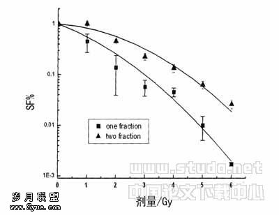
UⅡ以浓度[(10-9~10-7)mol/L]依赖性方式促进GMC 3H_TdR掺入,以UⅡ_4组刺激作用尤为明显。UⅡ 2~4组3H_TdR掺入量分别较对照组高86.9%,105.8%和134.2%(P<0.01)。而UⅡ_1组掺入量与对照组比无统计学意义(P>0.05,表1)。
表1 UⅡ对GMC 3H_TdR掺入的影响(略)
Table 1 Effect of UⅡ on 3H_TdR incorporation into GMC of rats
与对照组比 1)P<0.01
2.2 不同信号转导阻断剂对UⅡ作用的影响
UⅡ组3H_TdR掺入量较对照组高92.4%(P<0.01)。Ni、H7以及W7能明显抑制UⅡ 3H_TdR掺入,其抑制率分别为42.3%、49.3%和34.1%(P<0.01),PD98059不能够明显影响UⅡ的作用(P>0.05,表2)。
表2 细胞内信号转导阻断剂对UⅡ刺激GMC 3H_TdR掺入的影响(略)
Table 2 Effects of different signal transduction inhibitors on proli_feration of rats GMC induced by UⅡ
与对照组比 1)P<0.01;与UⅡ组比 2)P<0.01
3 讨论
UⅡ是继ET_1之后发现的又一新的缩血管活性肽,它能够收缩哺乳动物的大中动脉血管、气道和肠道平滑肌,收缩离体心肌组织,并有促丝裂作用,能刺激血管内皮细胞、平滑肌细胞、气道平滑肌细胞以及肿瘤细胞增殖,诱导心肌细胞肥大[9]。我们曾在离体孵育的大鼠主动脉组织上发现,UⅡ能以剂量依赖性方式激活NO合酶,尤其是血管固有型NO合酶,增加血管NO生成,促进血管外膜精氨酸转运[17,18]。我们曾报道慢性缺氧致肺动脉高压大鼠心肌UⅡ含量以及UⅡ受体结合位点明显升高[19],最近发现UⅡ能够协同异丙肾上腺素所致心肌肥大和心肌纤维化过程[20]。有学者报道肾功能衰竭患者和糖尿病肾功能衰竭患者UⅡ表达明显升高[21],提示UⅡ可能在肾脏功能稳态调节以及疾病发生中发挥重要作用,然而其作用机制尚不完全清楚。
本研究在体外培养的GMC上,采用3H_TdR掺入法,发现UⅡ能够以浓度依赖的方式促进细胞3H_TdR掺入增加,促进细胞增殖。该作用能够分别被Ca2+通道阻断剂NI、钙调素激酶阻断剂W7以及PKC抑制剂H7所阻断,提示凡影响胞浆Ca2+水平上的信号转导途径,即L_钙通道、CaM_PK以及PKC都能够影响对GMC的促增殖效应。然而,PD98059不影响UⅡ的效应,提示受体酪氨酸激酶_MAPK细胞增殖途径可能不是UⅡ促GMC增殖的主要途径。UⅡ的病理、生理意义及其作用机制是近年的研究热点之一。我们在国内首先报道了UⅡ的促丝裂效应。在培养的血管平滑肌细胞上,发现UⅡ能够促进细胞增殖,该作用需通过Ca2+、PKC、CaM_PK以及MAPK来实现[13]。我们还发现UⅡ能够促进新生乳鼠心肌成纤维细胞增殖和胶原分泌,其增殖效应与细胞外Ca2+内流、CaM_PK以及PKC激活有关,而促胶原合成作用可能通过其它途径来实现[22]。在慢性缺血所致肺动脉高压右心肥大大鼠上,我们发现右心室UⅡ以及受体结合位点明显上调,而血浆UⅡ水平并无明显变化,提出UⅡ可能主要以自分泌和旁分泌作用方式发挥效应这一观点[19]。上述结果进一步被其他学者所证实[4,9]。最近我们还发现UⅡ能够通过Ca2+、PKC以及MAPK途径刺激大鼠主动脉组织合成和释放内皮素_1[23]。一系列研究表明,UⅡ可通过GPR14受体、Ca2+、c_Src、PKC、MAPK和ROCK途径参与平滑肌细胞增殖以及促进巨噬细胞向泡沫细胞转化[9],并且MAPK途径激活还参与了UⅡ促进内皮细胞增殖的过程[14]。而本研究则证实UⅡ还能够通过激活L_钙通道、钙调素以及PKC途径刺激GMC的增殖。这与UⅡ促进心肌成纤维细胞的作用机制相近,但仍有待于进一步研究。
GMC是肾小球固有细胞,正常情况下GMC生长和分裂速度都很慢。但在病理情况下,生长和抑制失衡,GMC增殖活跃,产生大量细胞外基质,最终导致肾小球硬化和肾纤维化、肾功能进行性下降。许多因素能够促进GMC活化,使之发生表型转化并异常增殖,分泌细胞外基质和活性因子、炎性介质等,促使病变的发生和。本研究发现UⅡ能够促进GMC增殖,提示UⅡ可能在肾小球炎症与硬化等疾病的发病过程中发挥重要作用。
【】
[1]Coulouarn Y, Lihrmann I, Jegou S, et al. Cloning of the cDNA encoding the urotensin Ⅱ precursor in frog and human reveals intense expression of the urotensin Ⅱ gene in motoneurons of the spinal cord[J]. Proc Natl Acad Sci USA, 1998, 95 (26):15 803-15 808.
[2]Ames RS, Sarau HM, Chambers JK, et al. Human urotensin_Ⅱ is a potent vasoconstrictor and agonist for the orphan receptor GPR14[J]. Nature, 1999, 401(6 750): 282-286.
[3]Spinazzi R, Albertin G, Nico B, et al. Urotensin_Ⅱ and its receptor(UT_R)are expressed in rat brain endothelial cells, and urotensin_Ⅱ via UT_R stimulates angiogenesis in vivo and in vitro[J]. Int J Mol Med, 2006(6): 1 107-1 112.
[4]Watanabe T, Suguro T, Kanome T, et al. Human urotensin Ⅱ accelerates foam cell formation in human monocyte_derived macrophages[J]. Hypertension, 2005, 46(4): 738-744.
[5]Song W, Abdel_Razik AE, Lu W, et al. Urotensin Ⅱ and renal function in the rat[J]. Kidney Int, 2006, 69(8):1 360-1 368.
[6]Disa J, Floyd LE, Edwards RM, et al. Identification and characterization of binding sites for human urotensin_Ⅱ in Sprague_Dawley rat renal medulla using quantitative receptor autoradiography[J]. Peptides, 2006, 27(6): 1 532-1 537.
[7]Clozel M, Hess P, Qiu C, et al. The urotensin_Ⅱ receptor antagonist palosuran improves pancreatic and renal function in diabetic rats[J]. J Pharmacol Exp Ther, 2006, 316(3):1 115-1 121.
[8]Langham RG, Kelly DJ, Gow RM, et al. Increased expression of urotensin Ⅱ and urotensin Ⅱ receptor in human diabetic nephropathy[J]. Am J Kidney Dis, 2004, 44(5): 826-831.
[9]Watanabe T, Kanome T, Miyazaki A, et al. Human urotensin Ⅱ as a link between hypertension and coronary artery disease[J]. Hypertens Res, 2006,29(6): 375-387.
[10]Rakowski E, Hassan GS, Dhanak D, et al . A role for urotensin Ⅱ in restenosis following balloon angioplasty: use of a selective UT receptor blocker[J]. J Mol Cell Cardiol, 2005, 39(5): 785-791.
[11]Bousette N, Pottinger J, Ramli W, et al. Urotensin_Ⅱ receptor blockade with SB_611812 attenuates cardiac remodeling in experimental ischemic heart disease[J]. Peptides, 2006,27(11): 2 919-2 926.
[12]Gruson D, Rousseau MF, Ahn SA, et al. Circulating urotensin Ⅱ levels in moderate to severe congestive heart fai_lure: its relations with myocardial function and well established neurohormonal markers[J]. Peptides, 2006,27 (6): 1 527-1 531.
[13]张勇刚, 齐永芬, 夏春芳, 等. 尾加压素Ⅱ对血管平滑肌细胞增殖的影响[J]. 药通报, 2001, 17(2): 155-157.
[14]Shi L, Ding W, Li D, et al. Proliferation and anti_apoptotic effects of human urotensin Ⅱ on human endothelial cells[J]. Atherosclerosis, 2006, 188(2): 260-264.
[15]Johns DG, Ao Z, Naselsky D, et al. Urotensin_Ⅱ_mediated cardiomyocyte hypertrophy: effect of receptor antagonism and role of inflammatory mediators[J]. Naunyn Schmiedebergs Arch Pharmacol, 2004, 370(4): 238-250.
[16]Meger_Lehnert H, Tsai P, Caramelo C. ANF inhibites vasopressin induced Ca2+ mobilization and contraction in glome_ruler mesangial cells[J]. Am J Physio, 1988, 255, F771.
[17]姜志胜, 张勇刚, 齐永芬, 等. 尾加压素Ⅱ对大鼠主动脉一氧化氮合酶/ 一氧化氮系统的影响[J]. 北京大学学报(医学版), 2001, 33(2): 147-149.
[18]Lin L, Ding WH, Jiang W, et al. Urotensin_Ⅱ activates L_arginine/nitric oxide pathway in isolated rat aortic adventitia[J]. Peptides, 2004,25(11): 1 977-1 984.
[19]Zhang YG, Li JX, Cao J, et al. Effect of chronic hypoxia on contents of urotensin Ⅱ and its functional receptors in rat myocardium[J]. Heart Vessels, 2002,16(2): 64-68.
[20]Zhang YG, Li YG, Liu BG, et al. Urotensin Ⅱ accelerates cardiac fibrosis and hypertrophy of rats induced by isoproterenol[J]. Acta Pharmacol Sin, 2007, 28(1): 36-43.
[21]Totsune K, Takahashi K, Arihara Z, et al. Elevated plasma levels of immunoreactive urotensin Ⅱ and its increased urinary excretion in patients with Type 2 diabetes mellitus: association with progress of diabetic nephropathy[J]. Peptides, 2004 , 25(10): 1 809-1 814.
[22]张勇刚, 陈静炯, 许松. 尾加压素Ⅱ对大鼠心肌成纤维细胞增生和胶原合成的影响及其作用机制[J]. 中华老年心脑血管病杂志, 2001, 3(增刊): 41-43.
[23]张勇刚, 魏睿宏, 张?辉, 等. 尾加压素Ⅱ促进大鼠主动脉合成和分泌内皮素1的机制研究[J]. 中国动脉硬化杂志, 2007, 15(3): 165-168.